|
 Torrendiella dingleyae Torrendiella dingleyae
BiostatusPresent in region - Indigenous. Endemic
Images (click to enlarge)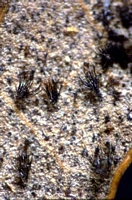
Owner: Peter Johnston | 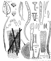
Caption: Fig. 7 A-G Torrendiella dingleyae (PDD 57727). A, Nothofagus solandri leaf with apothecia;
B, apothecia; C, seta; D, mature (right) and immature (left | 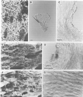
Caption: Fig.8 Torrendiella dingleyae (A-D,PDD64828;E-G,PDD66356). A, apothecia on leaf; B,
apothecium in vertical section; C, excipulum in vertical section; D, detail o | 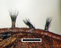
Caption: scale = 0.5mm
Owner: J.A. Cooper | |
Article: Johnston, P.R.; Gamundí, I.J. (2000). Torrendiella (Ascomycota, Helotiales) on Nothofagus. New Zealand Journal of Botany 38(3): 493-513 (http://www.rsnz.org/publish/abstracts.php).
Description: Apothecia developing on both sides of fallen leaves or on dead leaves
remaining attached to fallen twigs and branches; erumpent through host tissue, arising from
small patch of stromatic tissue at base of apothecium; apothecia 0.35-0.65 mm diam., short-stipitate, fleshy; disc plane, sepia grey or light greyish sepia; receptacle and stipe concolorous
with disc (darker in some collections), with numerous, black, stiff setae; stipe robust, short,
cylindric, often darkened near base. Paraphyses 1.5-2 µm diam., more or less
undifferentiated at rounded apex, about same length as asci. Asci 105-135 (-145) x 7.5-9 µm,
cylindric, tapering slightly to broadly subtruncate apex, wall thickened at apex with
prominent amyloid pore, 8-spored. Ascospores (11.5-) 12.5-14.5 (-15.5) x (4.5-) 5.5-6.5 (-7)
µm, broad ellipsoid-fusoid, tapering uniformly to each end, hyaline; uniseriate. Ectal
excipulum 3-layered; outer layer a single cell wide network of radially disposed, long-celled
hyphae 2.5-5 µm diam. with wall encrusted, dark brown, end cell often slightly swollen;
central layer comprising textura porrecta of long-cylindric cells with lumen 2.5-3 µm diam.
on sides of receptacle, 5-6.5 µm diam. near base, wall hyaline, thick, gelatinous; inner layer
comprising textura porrecta of long-cylindric cells 4-8 µm diam. with wall brown, thin,
nongelatinous. Setae arising from central layer of ectal excipulum, (300-) 400-500 (-600) x
12-15 (-20) µm, slightly swollen near the base, tapering to simple base, tapering gradually to
more or less acute apex, walls smooth, dark brown to black, slightly paler near apex,
pluriseptate (but septa visible only in pale upper portion of setae); setae on stipe shorter and
thinner than those on receptacle, about 190-230 µm long, tapering to either simple base, or
irregularly swollen at base, those with swollen base arising from outer layer of ectal
excipulum.
APPEARANCE IN CULTURE: OA: 80 mm diam.; aerial mycelium dense, grey, forming irregular
patches, lacking from most of colony; agar surface patchy with black, dark brown, and red
brown pigmentation. PDA: 35 mm diam., margin slightly uneven; colony surface near centre
raised and deeply convoluted; aerial mycelium low, dense, felted to cottony, whitish to pale
grey; in reverse very dark reddish brown. MEA-M: 75 mm diam.; aerial mycelium dense
felted to cottony, pale grey to pinkish; in reverse pale reddish brown, with irregular dark
brown patches. MEA-D: aerial mycelium sparse; agar with brown pigmentation, more intense
toward centre of colony, with small, scattered dark-brown flecks.
Notes: T. dingleyae can be distinguished macroscopically by its small, short-stipitate, dark
apothecia and numerous long, robust setae. Microscopically it can be distinguished from all
other species on Nothofagus by ascospore shape.
|