|
 Torrendiella cannibalensis Torrendiella cannibalensis
BiostatusPresent in region - Indigenous. Endemic
Images (click to enlarge)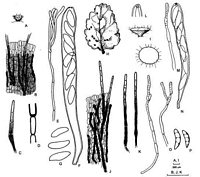
Caption: Fig. 3 A-G Torrendiella brevisetosa (PDD 64655). A, apothecium; B, surface view of the
receptacle; C, seta; D, detail of the base of a seta; E, 
Caption: Fig. 6 Torrendiella cannibalensis (A, PDD 57723; B-H, PDD 64242). A-B, apothecia in
vertical section; C, detail of B; D, excipular tissues near base of receptacle, in vertical | |
Article: Johnston, P.R.; Gamundí, I.J. (2000). Torrendiella (Ascomycota, Helotiales) on Nothofagus. New Zealand Journal of Botany 38(3): 493-513 (http://www.rsnz.org/publish/abstracts.php).
Description: Apothecia developing on both sides of fallen leaves; erumpent through host
epidermis, arising from small patch of compact tissue comprising angular cells with hyaline
walls, intermixed in part with host hypodermal cells; apothecia 0.3-0.8 mm diam., short-stipitate, fleshy; disc plane to slightly convex, translucent, greyish ("beige"), drying darker
("isabellinus"); receptacle slightly darker than hymenium, with reddish tint, with dark, stiff,
setae; stipe short, obconical, 0.5 mm long, 0.2-0.25 mm diam., concolorous with receptacle.
Paraphyses 1.5-3.5 µm diam., undifferentiated or slightly and irregularly swollen near
rounded apex, sometimes 1-branched, some containing yellowish guttules in upper part. Asci
(75-) 85-110 (-130) x (7.5-) 9.5-11.5 µm, cylindric to subclavate, tapering slightly to rounded
to subtruncate apex, wall slightly thickened at apex, pore with intense amyloid reaction, 8-spored. Ascospores (14.5-) 17-20 (-23) x 4-5 µm, elliptic, asymmetrical, flattened one side,
slightly curved, slightly wider in upper half, tapering to more or less acute apex, more
rounded toward base, hyaline; overlapping 1-2-seriate. Ectal excipulum 3-layered; outer layer
poorly developed, comprising loose network of hyphae 2-3.5 µm diam. with walls hyaline to
pale brown, with often numerous, short, irregular divarications, apical cells of hyphae often
with dense contents; central layer up to 40 µm thick, confined to stipe and base of receptacle,
on receptacle comprising regular rows of cylindric cells 2.5-4 µm diam. with walls hyaline,
thickened, gelatinous, this layer becoming partly lost and less well organised in older
apothecia, where it is confined to base of stipe, and comprises angular to subglobose cells
with walls hyaline, thickened, gelatinous; inner layer 10-30 µm thick, comprising rows of
long-cylindric cells 4-8 µm diam. with walls brown, thickly encrusted, nongelatinous. Setae
arise from central layer of ectal excipulum, (120-) 150-180 (-220) x 5.5-7.5 µm, swollen near
the base, tapering to base, tapering slightly to broadly rounded apex, walls smooth, brown but
not opaque (septa visible under microscope) in lower part, upper 3-4 cells pale brown to
hyaline.
APPEARANCE IN CULTURE: OA: 80 mm diam.; aerial mycelium sparse, in small scattered tufts
somewhat concentric in arrangement, pale grey; agar surface orange-brown; in reverse deep
orange-brown. PDA: 70 mm diam.; aerial mycelium dense, finely tufted, whitish; in reverse
deep reddish-brown. MEA-M: 50 mm diam.; aerial mycelium low, dense, crusty, whitish;
agar surface orange brown; in reverse deep orange brown; yellowish pigment diffusing into
agar across plate. MEA-D: 55 mm diam.; thin colony with sparse mycelium, pale brownish
pigment near centre.
Notes: T. cannibalensis is macroscopically distinct amongst the Nothofagus-inhabiting
species in possessing short-stipitate apothecia with a very pale receptacle, contrasting with
dark setae. T. eucalypti has a similar pale apothecium, but they are more distinctly stipitate,
and the setae are longer and less numerous. Excipular structure and appearance in culture
also differ between T. eucalypti and T. cannibalensis.
Two other Torrendiella species on Nothofagus in New Zealand have small,
macroscopically similar apothecia, T. brevisetosa and T. dingleyae. However, the ascospores
of T. brevisetosa are wider (6-7.5 µm), while the ascospores of T. dingleyae are symmetrical.
T. cannibalensis is also distinguished by its characteristic excipular structure; the outer layer
on the receptacle comprises hyphae with irregularly divaricating cells, while the central
gelatinous layer is confined mostly to the base of the apothecium. The excipulum comprises
mostly cylindric, brown-walled, nongelatinous cells, characteristic of the inner layer of the
ectal excipulum of Torrendiella.
|