|
 Torrendiella brevisetosa Torrendiella brevisetosa
BiostatusPresent in region - Indigenous. Endemic
Images (click to enlarge)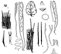
Caption: Fig. 3 A-G Torrendiella brevisetosa (PDD 64655). A, apothecium; B, surface view of the
receptacle; C, seta; D, detail of the base of a seta; E, 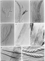
Caption: Fig. 4 Torrendiella brevisetosa (A-F, PDD 69778; GH, PDD 64655). A, apothecium in
vertical section; B, side of "typical" receptacle in vertical section with central gelatinous layer
of excipul | |
Article: Johnston, P.R.; Gamundí, I.J. (2000). Torrendiella (Ascomycota, Helotiales) on Nothofagus. New Zealand Journal of Botany 38(3): 493-513 (http://www.rsnz.org/publish/abstracts.php).
Description: Apothecia developing on both sides of fallen leaves, or on dead leaves still
attached to fallen branches; erumpent through host leaf epidermis, arising from small patch of
compact, hyaline textura intricata at base of stipe; apothecia 0.25-0.4 mm diam., sessile to
short stipitate, fleshy; disc plane, grey; receptacle dark grey, colour more intense at base, with
scattered, short, black, stiff setae; stipe short, broad, often paler than receptacle. Paraphyses
1.5-2 µm diam., slightly swollen to 3-4 µm diam. at rounded apex, branched, extending about
5 µm beyond asci. Asci (100-)115-140(-150) x (10-)12.5-14.5 µm, cylindric-clavate, apex
rounded to subtruncate, wall slightly thickened at apex, with indistinct, diffuse amyloid
reaction in outer part of wall, 8-spored. Ascospores (15-)17-20 x 6-7.5 (-8.5) µm, elliptic-fusoid, slightly wider in upper half, flattened on one side, often slightly curved, more or less
acute to the often slightly beaked apex, rounded to base, hyaline. Ectal excipulum varies in
appearance between apothecia; in some apothecia the layer of gelatinous tissue characteristic
of Torrendiella is well developed, while in others it is very poorly developed. In "typical"
apothecia outer layer poorly differentiated, comprising meandering hyphae 2 µm diam. with
walls finely encrusted and pale brown, forming sinuate pattern across surface of receptacle;
central layer up to 40 µm thick, comprising hyphae 2-2.5 µm diam. with walls thin, very pale
brown and finely encrusted, widely spaced within hyaline gelatinous matrix; inner layer 10-15
µm thick comprising textura porrecta of cylindric to brick-shaped cells 3-6 µm diam. with
walls brown, nongelatinous; at base of stipe, gelatinous tissue less well organised, forming
textura intricata of hyphae with walls hyaline, thick, gelatinous. In "atypical" apothecia,
gelatinous nature of tissue at sides of excipulum is lost, with the pale brown, finely encrusted
hyphae of central layer forming a complete, 1-3 cell wide layer across surface of receptacle;
inner layer remains same as in "typical" apothecia, as does gelatinous tissue at base of stipe.
Setae arising from central layer of ectal excipulum, 100-130 x 5-7.5 µm, often slightly
curved, inflated near the base, tapering to simple base, and gradually to rounded apex, walls
smooth, brown, slightly paler near apex, pluriseptate.
Notes: ETYMOLOGY: The specific epithet refers to the short setae characteristic of this species.
T. brevisetosa is characterised by its small, dark apothecia, relatively short, somewhat
curved setae, and large ascospores. T. dingleyae has similar small, dark apothecia, but it has
much longer setae, and differs in ascospore size and excipular structure.
T. brevisetosa is variable with respect to development of gelatinous tissue within the
excipulum. This difference appears to relate in part to maturity and aging of apothecia; all
immature apothecia (ascospores visible within asci but not fully differentiated) that have been
sectioned have a well-developed gelatinous excipulum, most mature apothecia (ascospores
fully differentiated within asci) lack the gelatinous layer. However, this is not always the case
in the genus, other species with poorly developed gelatinous layers (e.g., T. cannibalensis)
showing no variation in mature versus immature apothecia.
The type collection is from leaves that contain a mixture of T. brevisetosa and T.
cannibalensis apothecia. Although the two species sometimes occur adjacent on a single leaf,
in most cases the T. brevisetosa apothecia are on the upper surface of the leaves, the T.
cannibalensis apothecia on the lower surface. T. cannibalensis has narrower ascospores, and
an ascus pore with a more intense amyloid reaction.
A collection from fallen leaves of Nothofagus cunninghamii from Australia (Victoria,
Melba Gully, P. R. Johnston AU96-2 & B. Fuhrer, 17 May 1996, MEL) is macroscopically
similar in having small, dark apothecia with a pale stipe and short setae, a central excipular
layer in which the hyphae are held apart in a hyaline gelatinous matrix, and asci with a
similar, diffuse amyloid reaction at the apex (Fig. 5). The Australian species differs in having
slightly longer asci (140-160 µm), and slightly shorter and broader ascospores (14.5-16.5 x
6.5-8.5 µm) which are ovate and symmetrical in shape. Known from a single, mostly
immature collection, the species from Australia will not be formally described here.
|