|
 Sporisorium trachypogonis-plumosi Sporisorium trachypogonis-plumosi
BiostatusAbsent from region
Images (click to enlarge)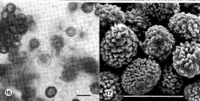
Caption: Fig. 16, 17. Spores and sterile cells of Sporisorium trachypogonis-plumosi (type) in LM
and SEM. Bars = 10 µm. | 
Caption: Fig. 18. Sori of Sporisorium trachypogonis-plumosi in some of the ovaries of
Trachypogon plumosus (type). Bar = 1 cm.
Fig. 19. Spore germination of Sporisorium trachypogonis-plumosi (type; on water-agar,
at room temp., in |
Article: Vánky, K. (1995). Taxonomical studies on Ustilaginales. XIII. Mycotaxon 56: 197-216.
Description: Sori (Fig. 18) in ovaries, long cylindrical, contorted or looped, 1.5-2 x 25-50 mm,
covered by an ashy-grey membrane which ruptures longitudinally to expose the granular-powdery mass of spore
balls and spores intermixed with irregular groups of sterile cells,
and a long, central, flagelliform columella. The distal part of the sori may be empty while
the proximal part may still contain immature, agglutinated spore masses. Spore balls
(Fig. 16) loose, irregular, many-spored, readily disintegrating. Spores (Figs. 16, 17)
globose, subglobose to broadly ellipsoidal, 6-8.5 x 7-10 µm, reddish-brown; wall 1-2.5
µm thick, finely tuberculate to coarsely echinulate (0.5-1.5 µm) under SEM, more
pronounced on the free surface of the peripheral spores. Sterile cells (Fig. 16)
subglobose, ovoid to slightly irregular, 5.5-11 µm long, thin-walled (ca. 0.5 µm), hyaline,
smooth, with 1-2 oil droplets. Spore germination (on WA, at room temp., in one day; Fig.
19) results in septate basidia which disintegrate readily producing lateral and terminal,
ellipsoidal basidiospores measuring 1-1.5 x 3-6.5 µm.
|