|
 Schizopora nothofagi Schizopora nothofagi
SynonymsPoria nothofagi
Hyphodontia nothofagi
Poria flavicans
Xylodon nothofagi
BiostatusPresent in region - Indigenous. Endemic
Images (click to enlarge)
Caption: Poria nothofagi x 600, spores x 1200. Part of dissepiment showing monomitic hyphal system with generative hyphae displaying clamp connections, capitate paraphysate hyphae, some crystal encrusted as are some ends of generative hyphae, and cylindrical spore
Owner: Cunningham, G.H. | 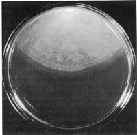
Caption: Fig. 7 Schizopora nothofagi (BAFC/cc 521). Aspect of the mat after 6 weeks of
growth. | 
Caption: Fig. 8 Schizopora nothofagi (BAFC/cc 521). Microscopic cultural characters: A,
generative hyphae; B, drepanocysts; C, allocysts. | 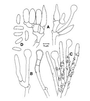
Caption: Fig. 13. Schizopora nothofagi. A, basidial clusters with cystidioles; B, bulbous hyphae; C,
encrusted hyphae from the dissepiments; D, spores. From the type of Poria nothofagi (PDD 5275). | |
Article: Cunningham, G.H. (1965). Polyporaceae of New Zealand. New Zealand Department of Scientific and Industrial Research, Bulletin 164: 304 p. Wellington:.
Description: Hymenophore annual, adherent, membranous, fragile, effused
forming linear areas 5-11 x 1-4 cm, sometimes extending to 1 metre, 1-3 mm
thick. Hymenial surface chalk white when fresh, drying cream or honey yellow, or
in parts pallid ochre, even, often creviced deeply; margin adherent, thinning
out, irregular, to 1 mm wide, finely fibrillose, cream. Pores not in strata,
irregular, orbicular, oval, angular, elliptical or labyrinthiform when slightly
irpiciform, 1-4 per mm, 0.2-1.5 mm diameter, to 1.5 mm deep, white or cream;
dissepiments fragile, 150-500 µm thick, irregular, edges bearing free capitate
paraphysate hyphae naked or encrusted with fine crystals. Context white or
cream, 0.1-1 mm thick, of intertwined hyphae encrusted with fine crystals;
generative hyphae 2.5-3 µm diameter, 0.25 µm thick, branched, septate, with
abundant clamp connections. Hymenial layer to 20 µm deep, a dense palisade of
basidia, paraphyses, and capitate paraphysate hyphae. Basidia subclavate, 12-14
x 3-4 µm, bearing 4 spores; sterigmata erect, to 6 µm long, commonly about 4 µm.
Paraphyses subclavate, 8-12 x 3-3.5 µm. Paraphysate hyphae projecting to 20 µm,
apices capitate, to 6 µm diameter, naked or bearing acecular crystals. Spores
mostly cylindrical, some suballantoid, apiculate, 5-6.5 x 1.5-2 µm walls smooth,
hyaline, 0.1 µm thick.
Habitat: HABITAT: Bark and decorticated wood of standing dead
saplings, fallen branches.and trunks, associated with a white rot.
Distribution: DISTRIBUTION: New Zealand.
Notes: Formerly described under the name of
Poria favicans (Karst.) G. H. Cunn. (Cunningham 1947b, p. 10), the
species has since been found to differ from this by its monomitic hyphal system,
absence of encrusted metuloids, much larger pores and fragile cretaceous
context. From related species of the section the plant differs in the capitate,
often encrusted paraphysate hyphae, which resemble those of Poria
versipora, and are abundant in the apices of dissepiments, cylindrical or
irregularly suballantoid spores, often labyrinthiform pores, fragile hymenophore
and restricted host range. When fresh plants are chalk white but when dry change
to some shade of yellow or ochre.
Article: Buchanan, P.K.; Ryvarden, L. (1988). Type studies in the Polyporaceae - 18. Species described by G.H. Cunningham. Mycotaxon 31(1): 1-38.
Notes: This characteristic species belongs in Schizopora Velen. emend. Donk and is readily separated from
the widespread S. paradoxa (Schrad.: Fr.) Donk by its cylindrical to suballantoid spores and
distinctly monomitic hyphal system. Otherwise, the two species have in common hyaline
cystidioles, bulbous hyphal ends, clamped, Hyphodontia-like hyphae, and coarse encrustation of
hyphae in the dissepiments. Spores of S. nothofagi measure 6-8(-10) x 2-3 µm, which is larger than
the measurements given by Cunningham (1965) of 5-6.5 x 1.5-2 µm.
Article: Rajchenberg, M. (1995). Notes on New Zealand polypores (Basidiomycetes) 2. Cultural and morphological studies of selected species. New Zealand Journal of Botany 33(1): 99-109 (http://www.rsnz.org/publish/abstracts.php).
Description: MACROSCOPIC CHARACTERS (Fig. 7): Growth very slow, 4.1-4.5 cm in 6 weeks.
Margin regular and subfelty. Mat poorly developed, pale cream, homogeneously
subfelty or pubescent with slightly raised mycelium. Around the inoculum denser,
white, and with inconspicuous radial stripes. Reverse bleaching. Odour none.
OXIDASE REACTIONS: gallic acid: +++, 0 cm; tannic acid: +, 0 cm; tyrosinase: +++, 0 cm
MICROSCOPIC CHARACTERS (Fig. 8): Generative hyphae with hemispheric clamps,
hyaline, slightly thickened walls, 3-5 µm diam., branched, also with short digitiform
lateral branches 1-1.2 µm diam. Drepanocysts present from the 4th week onwards.
Allocysts are formed in the mat around the inoculum.
SEXUALITY: Tetrapolar. Ten single spore cultures were confronted with each other
and were distributed as follows: BAFC 521: A1B1: 1, 4, 8, 10; A2B2: 9;
A1B2: 2, 6; A2B2: 3, 5, 7;
MR 10057: A1B1: 1, 10; A2B2: 9, 8; A1B2: 3, 5, 7;
A2B1: 6, 11
NUCLEAR BEHAVIOUR: Normal.
CODE: 2.3c.7.26.31 b.32.36.40.47.54.60.61
Notes: REMARKS: The formation of drepanocysts in culture stresses once again the close
relationship between the polyporoid Schizopora and the corticioid Hyphodontia .
Erikss. (Donk 1967; Hassan Kassim & David 1983; Eriksson et al. 1984; Gilbertson &
Ryvarden 1987).
|