|
 Rosellinia mammoidea Rosellinia mammoidea
SynonymsPsilosphaeria mammoidea
BiostatusPresent in region - Indigenous. Endemic
Images (click to enlarge)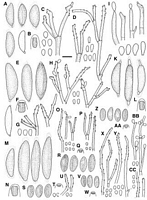
Caption: Fig. 12. S-U, R. mamoidea, type, K 308: S, Ascospores (spore on right PDD 71726); T, Ascus apical ring; U, Conidiophores and conidia in culture (PDD 71726). Scale bar = 10 µm. | 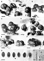
Caption: Fig. 20 Rosellinia mammoidea. A-F, Stromata, A with remnants of subiculum (arrow); G,
Ascospores, 3rd showing cellular appendage (arrow), 4th to 6th showing short germ slit; H,
| 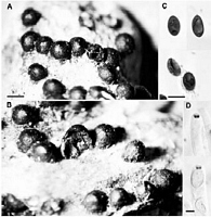
Caption: Fig. 19 Rosellinia mammoidea. A, B, Stromata;
C, Ascospores; D, Ascus apical rings in
Melzer's reagent. Type, K 308. Scale bars: A, B = 0.5 mm; C = 10 µm; D = |
Article: Petrini, L.E. (2003). Rosellinia and related genera in New Zealand. New Zealand Journal of Botany 41(1): 71-138 (http://www.rsnz.org/publish/abstracts.php).
Description: Subiculum evanescent, white, cream to light brown to grey. Stromata (350)482 ± 80(650) µm
high, (500)658 ± 105(900) µm wide (n = 25), cylindrical to semiglobose, black, shiny, solitary
or crowded in small groups, rarely 2-3 fused together. Ostioles finely papillate. Ectostroma
50-75 µm thick, black. Entostroma not seen. Perithecia detached in mature material. Ascus
apical rings 2-3 µm high, upper width 2.8-4 µm, lower width 1.9-3 µm (n = 17), without
bulge at upper margin, J+, blue. Ascospores (11)13 ± 1(16) µm long, (6.2)7.5 ± 0.5(9) µm
wide (n = 150), inequilaterally ellipsoidal, brown to dark brown, with 8-10 µm long straight
germ slit (Fig. 20G), some of them with a basal, 1 x 1 µm large, cellular appendage (Fig.
20G).
Culture on MA white, felty, grey areas with conidiophores. Conidia 3.5-5 x 3-4 µm.
ANAMORPH: Geniculosporium.
Habitat: HOSTS: Metrosideros robusta, unidentified dicotyledonous wood.
MATRIX: Decorticated heavily decomposed wood.
Notes: NOTES: Rosellinia mammoidea is characterised by a cream to light brown subiculum present
only in a very early state, and dark brown ascospores with rounded side walls with a germ slit
about two thirds of their length. In the original description the ascospore size ranges from 16
to 18 x 8 µm (Cooke 1879). The Kew herbarium has three specimens labelled as R.
mammoidea from the period when Cooke described the fungi from New Zealand (Cooke
1879). One originates from Wellington, collected by Travers, the second from the South
Island, J. Kirk 72, the third from Waitaki, ex herb. M. C. Cooke. The Travers collection is
cited by Cooke and is labelled as the type. The ascospore size in this specimen ranges from 11
to 14 x 6.5 to 8 µm, clearly much smaller than the dimensions given in the literature. The Kirk
specimen is R. communis: its ascospores measure 16-21 x 8.5-10 µm. The third specimen has
ascospores measuring 19-23 x 10-13 µm with a sigmoid germslit and is Helicogermslita
aucklandica. At first sight, all three have similarstromata.
The wrong ascospore size indicated in the literature was very likely the reason why
most Rosellinia from New Zealand identified as R. mammoidea are actually R. communis.
Rosellinia mammoidea can be distinguished from R. communis by smaller stromata and
smaller ascospores, and from R. johnstonii by smaller stromata with mostly rounded tops,
larger (usually wider) ascospores, occasionally with a cellular appendage and shorter germ
slits positioned symmetrically. The results of the discriminant analysis of the ascospore size
indicated statistically significant differences among these three species, as also shown by the
65% confidence ellipses in Fig. 9C. The stromatal size was also statistically
significantly different (data not shown). R. mammoidea differs also from R. subiculata
(Schwein. : Fr.) Sacc. by the subiculum colour, larger ascospores, and a shorter germ slit
(Petrini 1993).
Martin (1968) treated R. mammoidea as a synonym of Hypoxylon mastoideum (Fr.)
P.M.D.Martin ( Rosellinia mastoidea (Fr.) Sacc.) and gave its spore size as 10-22 x 5-10 µm.
Such a large variability in ascospore size is most likely the result of including more than one
taxon in the species concept. According to Petrini (1993) its basionym, Sphaeria mastoidea
Fr., remains doubtful. Martin (1968) drew his taxonomic conclusions mainly from material
collected in South Africa. Based on my experience, the geographical distribution of most
species of Rosellinia is restricted. Therefore, the New Zealand material, which originates from
an isolated area, is almost certainly different from Sphaeria mastoidea which very likely
originates from Europe. South African material still needs to be studied in order to establish
its identity.
Article: Cooke, M.C. (1879). New Zealand fungi. Grevillea 8(46): 54-68.
|