|
Chlorovibrissea phialophora
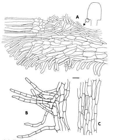
Caption: Fig. 3. Chlorovibrissea phialophora. A, Margin with portion of hymenium (on left) and ectal excipulum (on right). B, Hairs from surface of stipe. C, Medullary tissue of stipe. All drawn from the holotype. Scale bar = 10 µm.
|