|
 Mycena mariae Mycena mariae
BiostatusPresent in region - Indigenous. Endemic
Images (click to enlarge)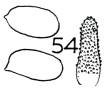 | 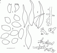
Caption: Fig. 8 M. mariae. 1. basidia. 2. cheilocystidia. 3. basidiospores. 4. pleurocystidium.5. pileipellis elements. 6a. caulocystidia. 6b. terminal cells of the stipe (PDD 56757). | 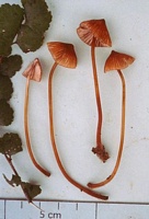
Owner: Karl Soop | 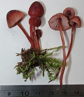
Caption: fruitbody
Owner: J.A. Cooper | 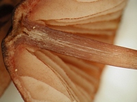
Caption: gills showing dark edge
Owner: J.A. Cooper | 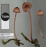
Caption: FUNNZ photo
Owner: J.A. Cooper | 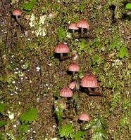
Caption: FUNNZ photo
Owner: J.A. Cooper | 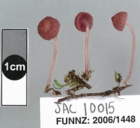
Caption: FUNNZ photo
Owner: J.A. Cooper | 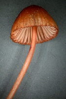
Owner: J.A. Cooper | 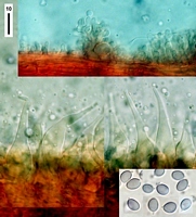
Caption: top: cap ornamented hyphae. Middle: cheilocystidia. Bottom right: spores.
Owner: J.A. Cooper | 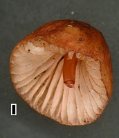
Caption: scale = 1mm
Owner: J.A. Cooper | 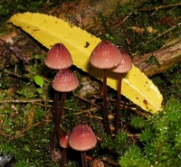
Owner: J.A. Cooper | 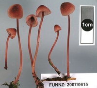
Owner: J.A. Cooper | 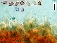
Caption: cheilocystidia and spores
Owner: J.A. Cooper | 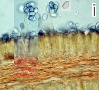
Caption: pleurocystidia and section through trama (in melzers)
Owner: J.A. Cooper | 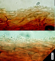
Caption: section through cap
Owner: J.A. Cooper | 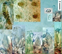
Caption: tope left & centre: pleurocystidia. Bottom left & centre: cheilocystidia. Bottom right & top: basidia and spores.
Owner: J.A. Cooper | 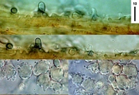
Caption: cap surface hyphae. Lower: cap cells?
Owner: J.A. Cooper | 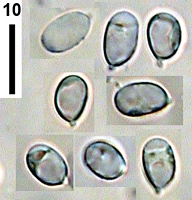
Caption: spores
Owner: J.A. Cooper | 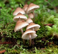
Owner: P. Leonard | 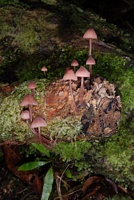
Owner: J.A. Cooper | 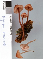
Owner: J.A. Cooper | 
Caption: FUNNZ2007/0615
Owner: FUNNZ | 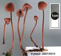
Caption: FUNNZ2007/0615
Owner: FUNNZ | |
Article: Segedin, B.P. (1991). Studies in the Agaricales of New Zealand: some Mycena species in sections Longisetae, Polyadelpha, Rubromarginatae, Galactopoda, Lactipedes, and Calodontes. New Zealand Journal of Botany 29(1): 43-62 (http://www.rsnz.org/publish/abstracts.php).
Description: Microscopic characters of the holotype
Spores 8.5-11.5 X 5.5-7 (9.1 X 6.3) µm., Q=1.4, distinctly
elliptical to elliptic elongate, variable in size, collapsing-easily, strongly
amyloid, walls becoming eroded during drying. Cheilocystidia 50-65 X 9-13 µm.,
narrow fusoid, fusoid-ventricose to lageniform, some sharply pointed apically,
yellow-brown in KOH. Pleurocystidia like cheilocystidia, infrequent. Basidia
short and fat (20 X 10 µm.) with 2 or 4 plump sterigmata. Trama of inflated
cells (-15 µm. diam.), dextrinoid in Melzer's, with numerous lactifers, narrow
hyphae with dark red (in KOH) contents. Subhymenium a narrow zone of narrow
hyphae, strongly dextrinoid in Melzer's. Pileipellis narrow, repent hyphae with
narrow, mostly short, simple protuberances, not easy to determine. Sub-pellis
of large, inflated cells up to 30 µm. diam. Context of narrow and inflated hyphae
5-15 µm. diam. Stipe of parallel, narrow, thick-walled hyphae, dark coloured;
some simple or diverticulate protuberances, especially towards the base. Clamp
connections seen occasionally. Dried material dark red to black.
The type material
appears to be a mixed collection. There are two pale brown stipes, one attached
to wood, which do not seem to be related to the rest of the material. They bear
a quantity of brown spore on their surface, rein forcing the impression that
they belong to some other fungus. The rest of the material although somewhat
fragmented, adds up to two fruiting bodies, as illustrated in Stevenson's painting
and these provided the information above Stevenson's drawing of a cheilocystidium
of M. mariae is still a puzzle for it is of the Rotalis type which one
would not expect in section Galactopoda It could perhaps have been a drawing
of a pileipellis element, and there is still the possibility that the collection
is a mixed one.
Description of basidiome based on information from a further collection.
Pileus 14 X 6 mm.,
dull pinkish red, convex, broadly umbonate, smooth to Finely fibrillose. Lamella
ascending, adnate with decurrent tooth, 2 series, up to 16 reaching the stipe,
pink with a red margin. Stipe 30-40 X 1 mm, concolorous with pileus, hollow
expanding slightly towards the base, paler at to darker below, exuding red latex
when broken, long brown hairs at the base. Flesh thin, pink in cap, darker in
stipe. Smell and taste unknown. Fungus drying dark red to black. The red colour
in the basidiome dissolves out readily in KOH and stains the cytoplasm of spores
umber.
Microscopically
PDD 56757 fits very well with the description of the type, the main variation
being in the greater degree of diverticulation in the pileipellis elements and
more distinctly diverticulate terminal cells on the stipe (Fig. 8: 6b). The
spores were also slightly longer (8.5-12.5 X 5.5-7 (10.7 X 5.9) µm, Q= 1.8).
Habitat: HABITAT: In litter in mixed podocarp-dicotyledonous
forest.
Notes: Distinguishing
features of M. mariae are the large, elongate spores and the dark red
pigment, which exudes into the mounting paper and into mounting medium. It is
quite distinct from M. morris-jonesii (see description below) by virtue
of its colour and shape and size of spores. The tissues of M. mariae
are noticeably more dextrinoid than those of M. morris-jonesii. There
does not seem any doubt that these are different species and it is therefore
proposed that the Stevenson species, M. mariae Stevenson, be reinstated.
Article: Stevenson, G. (1964). The Agaricales of New Zealand: V. Kew Bulletin 19(1): 1-59.
Description: Pileus 1.5-2 cm. diam., dark testaceous, campanulate, more or less umbonate, sub-fibrillose; flesh moderately thin, whitish. Gills sinuately adnexed, whitish with dull red margins and some red blotches, moderately crowded. Stipe 5-6 cm x 2-4 mm, testaceous to pale testaceous, smooth with spreading hyphal hairs at base, producing some red latex when broken. Spores 6 x 11 µm, amyloid, thin-walled. Hymenophoral trama and tissue of Pileus pseudo-amyloid. Cheilocystidia 25-30 x 10 µm, ornamented (Fig. 54).
Habitat: In forest litter, Woodside, Dunedin, 23.5.1953, Stevenson (type). Named after Mrs. Marie Taylor.
Article: Horak, E. (1971). A contribution towards the revision of the Agaricales (Fungi) from New Zealand. New Zealand Journal of Botany 9(3): 403-462 (http://www.rsnz.org/publish/abstracts.php).
Notes: Mycena mariae Stevenson (29 D) Fig. 15 = Galactopus morris-jonesii
(Stevenson) Horak
Spores oval, hyaline, amyloid, smooth, 8-11.5 X 5-6 µ. Cheilo- and
pleurocystidia fusoid or awl-shaped, thin-walled, filled with a reddish cell sap,
55-75 X 10-16 µ.
|