|
 Mycena globuliformis Mycena globuliformis
BiostatusPresent in region - Indigenous. Endemic
Images (click to enlarge)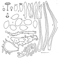
Caption: Fig. 1 M. globuliformis. I. basidiomes. 2. basidia. 3. cheilocystidia. 4. basidiospores. 5. pileipellis elements. 6. subpellis elements. 7. caulocystidia. |
Article: Segedin, B.P. (1991). Studies in the Agaricales of New Zealand: some Mycena species in sections Longisetae, Polyadelpha, Rubromarginatae, Galactopoda, Lactipedes, and Calodontes. New Zealand Journal of Botany 29(1): 43-62 (http://www.rsnz.org/publish/abstracts.php).
Description: Pileus up to 14
mm diam., almost spherical, resting on the basal disc, when young; as the stipe
elongates it becomes convex to shallow umbilicate when older, sulcate, pellucid
striate at margin, straw-coloured to darker greenish-yellow in depressed centre;
greenish cottony flecks over surface when young, confined to depressed centre
when older; distinctly viscid, probably with a separable pellicle. Lamellae
free, apparently not attached to a collar, creamy white with a darker, strongly
fimbriate edge, thin, crowded, 2 series, 20 reaching the stipe. Stipe up to
15 X 1-2 mm, even above a conspicuous basal disc, white at top, slightly yellow
towards the base, densely covered with caulocystidia (villose), hollow; disc
lined with a mat of hairs and with a black margin and under surface in most
specimens. Stipe breaks off readily at the base and the disc persists, attached
to the substratum, for some time. Smell and taste unknown.
Colour of spore print unknown.
Spores 8-11 X 5.5-7
(9.4 X 6.3) µm., Q = 1.49, ellipsoid, hyaline, thin-walled, strongly amyloid.
Basidia 15-18 X 6-8.5 µm., short and fat, 2- to 4-spored, sterigmata relatively
long (up to 4-5 µm.). Cheilocystidia 35-40 X 3-7 µm., forming a broad band of
closely packed, thin-walled, fusiform to narrowly clavate cells, mostly with
a smooth apex but sometimes with 1-4 µm. small apical protuberances, yellow-brown
plasmatic pigment. Gill edge gelatinised. Pleurocystidia absent. Trama of more
or less parallel, narrow (2-3 µm.) hyphae, vinescent-red in Melzer's. Context
of thick-walled hyphae of fairly regular cells (15 X 7 µm.), not inflated, vinescent-red
in Melzer's. Pileipellis very complex; suprapellis of narrow, gelatinised hyphae,
probably separable, with scattered, large bundles of thin-walled hairs; pellis
of narrow (5-6 µm.) hyphae with short, sometimes diverticulate protuberances
often terminating in inflated cells (10-15 µm. diam.) bearing large numbers
of short (1-4 µm.) to long (-10 µm.) protuberances. Beneath the pellis is a
subpellis of mostly spherical (4.5-5 µm. diam.) to short cylindrical (9-16 µm.
long) elements with very thick (up to 3 µm. wide) walls. Stipe of narrow hyphae,
the cortex bearing many long, thick-walled, tapering caulocystidia (85-140 X
6-12 µm. at the base, 2-4 µm. diam. at the tips, some distinctly swollen at
the base). In the middle of the stipe are a few conducting hyphae with yellow
(KOH), shining contents. Disc consists of thick-walled, hyaline hyphae 4 µm.
diam., with a brown, resinous, encrusting pigment; the dark colour at the edge
and underside of the disc appears to be due in part to colonies of blue-green
algae. No clamp connections seen.
Habitat: HABITAT: Gregarious, on dead wood in mixed podocarp-dicotyledonous
forest.
Notes: ETYMOLOGY: The epithet of the new species reflects the
almost completely spherical form of the young basidiome.
At first sight,
the assigning of this fungus to any section as presently defined raises some
problems. The well-developed disc at the base of the stipe and gelatinous pellicle
suggest section Basipedes but it would be excluded on account of its green colour
(Maas Geesteranus 1980). Maas Geesteranus has not included section Cyanocephalae
in his Conspectus, presumably because it comprises only Southern Hemisphere
species. This section was introduced by Singer in 1975 but not legitimised until
1986 (Singer 1986), to accommodate three blue to blue-green species; M. cyanocephala
Sing., (S. America) M. interrupta (Berk.) Sacc. (Australia), and M.
veneta Stev. (New Zealand), all of which Horak (1983) considers to be the
same species, namely M. interrupta.
M. globuliformis can be confused with M. interrupta, even when it is found on
the same log (PDD 34786), but, when looked at critically, the pileus is seen
to be more yellowish-green on a straw-coloured background, rather than blue-green,
which would exclude it from section Cyanocephalae. The situation is further
confused by the presence of blue-green algae at the edge of the disc, which,
when moist, may give a distinctly blue-green colour. The appropriate place for
M. globuliformis appears to be section Longisetae, despite the absence
of pileal setae and the amyloidity of the spores. There are, indeed, many resemblances
to M. longiseta Hohnel, such as structure of the pileipellis, the cheilocystidia
and the disc, and the long setiform hairs on the stipe. Although there are no
pileal setae, the subpellis in M. globuliformis is composed of conspicuously
thick-walled cells very much like the basal cells, which are extended into setae
in M. longiseta. Another species of Mycena, M. sublongisetae
Z.-s. Bi, has recently been described from China. This differs from M. globuliformis
in its larger size, its yellow-brown pileus and orange-yellow lamellae, but
it resembles it in having slightly amyloid spores; the pileal setae are intermediate
in size between the other two species. The discovery of a third species related
to M. longiseta reinforces the segregation of section Longisetae from
section Basipedes.
|