|
 Cheilymenia catenipila Cheilymenia catenipila
BiostatusPresent in region - Indigenous. Endemic
Images (click to enlarge)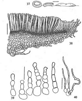
Caption: Figs 37-40. Cheilymenia calenipila J. Moravec sp.nov. 37 -revived (rehydrated) apothecia
(scale bar =1 mm); 38 - median section through apothecium (showing cyanophilic cells of
hypothecium and ectal excipulum stained with C413) (scale bar = 100 | 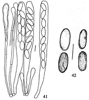
Caption: Figs 41-42. Cheilymenia catenipila J. Moravec sp.nov.: 41 - paraphyses and asci (scale bar =
10 µm); 42 - ascospores (oil immersion, C4B, scale bar =10 µm). Holotype (WELTU 57). | 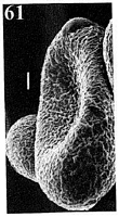
Caption: Fig 61. SEM photomicrographs of ascospores of Cheilymenia: 61 C. catenipila J. Moravec sp.nov.
(holotype WELTU). Scale bars = 1 µm. | 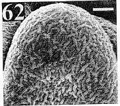
Caption: Fig 62. SEM photomicrographs of ascospores of Cheilymenia: 61 C. catenipila J. Moravec sp.nov.
(holotype WELTU). Scale bars = 1 µm. | |
Article: Moravec, J. (2003). A taxonomic revision of the genus Cheilymenia Boud. - 9. The sections Villosae and Obtusipilosae, and a revision of the genus Pseudoaleuria Lusk (Pezizales, Pyronemataceae). Acta Musei Moraviae. Scientiae Biologicae 88: 37-73 Brno:.
Description: Apothecia (Fig. 37) small to medium sized, 1.5-3.5 mm in diam., first subglobose, becoming
shortly dolfform, then shallowly saucer-shaped and finally applanate to moderately pulvinate;
hymenium pale orange, receptacular surface slightly paler, minutely granulate, faintly
fimbriate at margin with protruding, very short, subhyaline hairs only sparsely distributed on
the receptacular surface.
Apothecial structure (Fig. 38). Hymenium about 140-165 µm thick. Hypothecium about 20-45 µm thick,
differentiated from medulla as consisting of much smaller, mostly rounded,
densely-packed cyauophilic cells 3-8 µm in diam.; medulla about 70-90(-130) µm thick, of a
textura angularis to subintricata, consisting of irregularly angular cells 8-25 µm in- diam.
occasionally mixed with septate, 6-10 µm thick hyphae. Ectal Excpulum about 45-80(-120)
µm thick, becoming thinner towards margin, of a textura globulosa to subangularis composed
of large subglobose, rarely subangular, yellowish, strongly cyanophilic cells 10-40 µm in
diam; towards the margin the cells become elongate-clavate forming marginal rim together
with protruding catenulate hairs.
Hairs (Figs 39) short, 25-50 x 6-12 µm, originating from globose cells of ectal excipular
layer, comparatively sparsely distributed on whole receptacular surface; both marginal and
lateral hairs are of similar size and shape, mostly catenulate, consisting of hyaline or
subhyaline globose or elongate cells or subcylindrical articles, rarely with continuous walls
and septate, thin-walled, each hair terminating in globose or often elongate or pyriform cell
with irregularly thickened and often yellow-brownish darkened wall (0.1-0.7 µm). Subicular
hyphae (Fig. 40) occur at the apothecial base; they -are hyaline, flexuous, interwoven, mostly
thin-walled (walls 0.1-0.3(-0.8) gm thick). Asci (Fig. 41) 140-160x10-13 µm, widely
cylindrical, with rounded or mostly moderately constricted and subtruncate apex, gradually
constricted towards simple or bilobed base, eight-spored. Ascospores (Figs 42, 61-62)
uniseriate or mostly biseriate, narrowly ellipsoid, often very narrow, occasionally
subcylindrical, rarely wider, (9.5-)10.2-13.0(-14.5) x (4.3-) 4.7-5.8(-6.9) µm, (mostly 12.0 x
5.5 µm), hyaline; at maturity possessing a feeble yellow refractive colour when stained with
C4B; loosening perispore nearly smooth, densely and irregularly covered with extremely fine
cyanophilic ornamentation which is clearly recognisable only on SEM photomicrographs
(Fig.) as densely arranged, blunt, vermicular, fine crests. Paraphyses (Fig. 41) filiform,
1.7-2.7 µm thick, straight, sparsely. septate, apices clavate-dilated to 4.0-7.5(-8.0) µm.
Habitat: Coprophilous, on cow dung.
Distribution: Known only from the type locality in
New Zealand.
Notes: Etymology. Derived from the Latin catenulatus (catenulate, arranged in series of chains) and
pilus (hair), referring to the hairs, which are composed of cells that are mostly arranged in
chains.
Remarks. C. catenipila is rendered a rather peculiar species by the catenulate shape of the
hairs. However, some hairs are articulate-septate and closely resemble hairs of C. magnifica.
The new species probably indicates a further link to the genus Pseudombrophila Boud., but
the absence of a dark brown or violaceous-purple amorphous pigment in the excipulum, in
the hairs and paraphyses, as well as the clavate shape of the paraphyses and the loosening
ascospore perispore, confirm the classification in Cheilymenia. The shape of the apothecia
makes it easily confused with a species of the section Coprobia. However, as mentioned
above, all species of the section Coprobia (Boud.) J. Moravec (1990b) differ in having only
hyphoid hairs, and ascospore perispore covered with distinct longitudinal rib-like striation.
Thus, like other genera of Pyronemataceae, Cheilymenia also does not appear to be a sharply
delimited genus but, obviously, several phylogenetic links are indicated. As already
mentioned, the sect. Obtusipilosae is very close to the genus Pseudoaleuria Lusk, which has
been revised and is addressed below.
|